

5. Development of the extraocular motor system
Eye movements require dynamic specializations of extraocular muscle fibers and their innervation. We focus on the morphological organization of eye muscles and their motor innervation in larval Xenopus. This study employs various immunostainings for muscle and nerve fiber types, confocal laser scanning of whole eye muscles and semi-thin sections of embryonic and larval Xenopus. Comparing different stages of Xenopus allows a detailed analysis of the developmental plasticity.
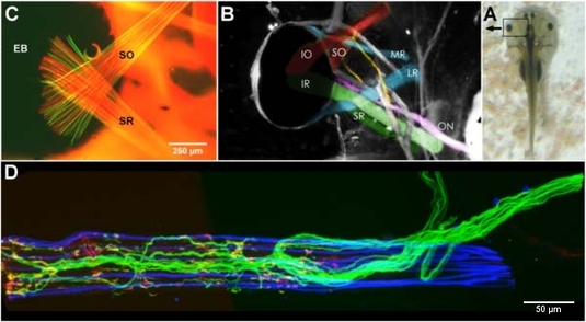
A - Xenopus larva before metamorphosis.B - Colored overview of the extracular muscles in Xenopus tadpole.C - Fluorescent stainings for f-actin (red, phalloidine) and slow myosin (green, MHCs-antibody), insertion site on eye bulb shown.D - Confocal scanned inferior rectus at stage 47 with triple fluorescence labelling of slow myosins (blue, MHCs-antibody), motor nerve (green, dextran tracer) and motor endplates (red, bungarotoxin).
Back to Research group: Sensory motor transformation